Diag image, short for diagnostic imaging, is a branch of medicine that uses advanced imaging technologies to capture pictures of the inside of the body. These images allow doctors to observe organs, tissues, and structures non-invasively, helping them make accurate decisions about health conditions. From detecting broken bones to identifying tumors, diag image plays a vital role in modern healthcare.
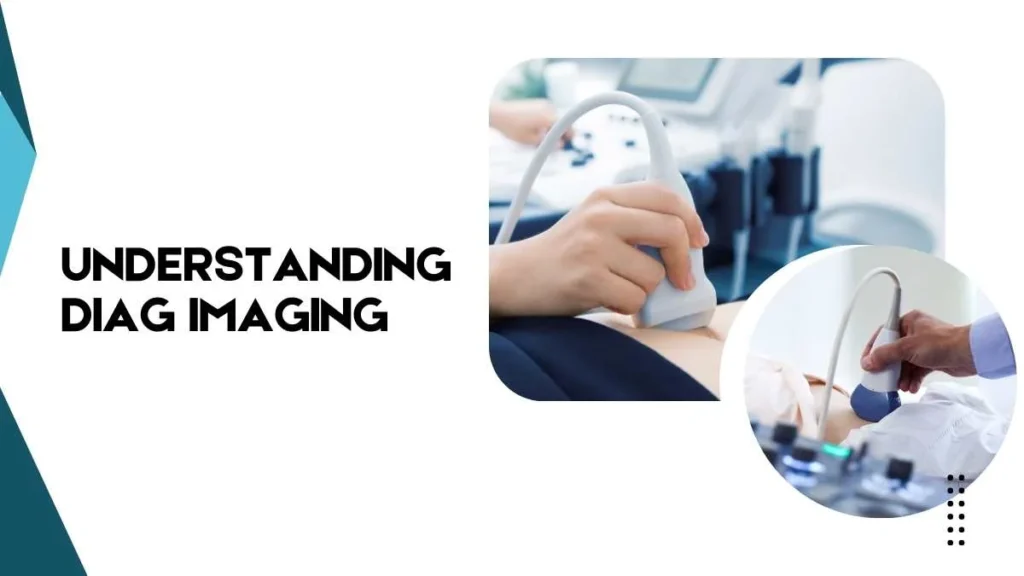
In simple words, diag imaging refers to a set of medical imaging techniques such as X-rays, CT scans, MRIs, ultrasounds, and PET scans. These tools help medical professionals diagnose, monitor, and sometimes even guide treatment without the need for surgical procedures. Today, diagnostic imaging is considered one of the most powerful tools in preventive medicine and accurate disease management.
Historical Background
The roots of diagnostic imaging go back to 1895, when Wilhelm Conrad Roentgen discovered X-rays. This discovery marked the first time doctors could see inside the body without surgery.
Milestones since then include:
- 1920s–30s: Use of contrast agents to enhance imaging detail.
- 1950s: Development of ultrasound technology, especially in obstetrics.
- 1970s: Introduction of Computed Tomography (CT), offering cross-sectional imaging.
- 1980s: Clinical adoption of Magnetic Resonance Imaging (MRI).
- 2000s: Growth of hybrid imaging, such as PET-CT and PET-MRI.
Evolution of Diagnostic Imaging
Diagnostic imaging has revolutionized healthcare. In the past, diagnosing internal conditions often required exploratory surgery. With the invention of X-rays in 1895, the medical field entered a new era. Since then, innovations like CT scans, MRI, and ultrasound have made it possible to visualize nearly every part of the human body in detail.
Each advancement has improved accuracy, safety, and speed, making diagnostic imaging an essential pillar of evidence-based medicine. It not only helps doctors diagnose conditions but also supports ongoing monitoring and effective treatment planning.
Key Types of Diagnostic Imaging
Here are the most common diag image techniques used in healthcare:
| Type of Imaging | How It Works | Common Uses |
| X-ray | Uses electromagnetic radiation to capture bone and organ images | Detect fractures, chest conditions, and dental exams |
| CT Scan | Combines X-rays with computer processing to create 3D images | Detect cancers, internal bleeding, and organ damage |
| MRI | Uses magnetic fields and radio waves for detailed images | Brain scans, spinal cord injuries, joint problems |
| Ultrasound | Uses sound waves to produce images in real-time | Pregnancy scans, organ health, and blood flow studies |
| PET Scan | Uses radioactive tracers to assess cellular activity | Cancer detection, heart disease, and brain disorders |
Importance of Diag Image in Healthcare
Diag image is indispensable in medicine because it provides clarity when symptoms are vague or invisible. For example:
Early Detection:
Cancers, heart diseases, and neurological disorders can be diagnosed in their early stages.
Accurate Monitoring:
Doctors can monitor the progress of treatment, such as chemotherapy or surgery recovery.
Non-Invasive Insight:
Patients avoid unnecessary surgical procedures, reducing risk and discomfort.
Emergency Care:
In trauma cases, imaging quickly identifies internal injuries that are otherwise undetectable.
By offering real-time insights, diagnostic imaging often makes the difference between life-saving interventions and delayed treatment.
Common Applications of Diag Imaging
Diag image techniques are applied across nearly every medical specialty:
- Oncology: Detecting tumors and tracking cancer treatment.
- Cardiology: Monitoring heart function and identifying blockages.
- Neurology: Mapping brain activity and diagnosing strokes or multiple sclerosis.
- Orthopedics: Identifying fractures, arthritis, or joint issues.
- Obstetrics and Gynecology: Monitoring pregnancies and reproductive health.
Safety and Risks in Diagnostic Imaging
While diagnostic imaging is generally safe, certain procedures involve risks. For example, X-rays and CT scans expose patients to radiation, though modern technology has significantly minimized exposure levels. MRI scans are safe but may be unsuitable for patients with pacemakers or metal implants. PET scans involve radioactive tracers but are carefully monitored to ensure safety.
Healthcare providers weigh the benefits against risks, ensuring that diagnostic imaging is used responsibly. In most cases, the advantages far outweigh the minimal risks.
Future of Diagnostic Imaging
The future of diag image is promising, with rapid advances in technology. Artificial intelligence (AI) is increasingly being used to analyze scans, detect patterns, and even predict diseases earlier than human eyes can. Portable imaging devices are making diagnosis more accessible in rural areas. Additionally, hybrid imaging techniques like PET-MRI combine the strengths of multiple methods for unmatched precision.
These innovations mean that diagnostic imaging will continue to improve accuracy, accessibility, and patient outcomes.
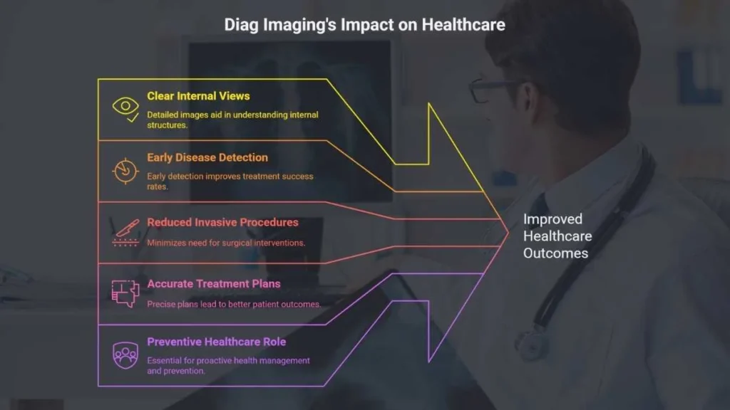
Benefits at a Glance
- Provides clear, detailed views of internal structures
- Enables early disease detection
- Reduces the need for invasive procedures
- Enhances the accuracy of treatment plans
- Plays a critical role in preventive healthcare
FAQs
Q: Is diagnostic imaging painful?
No, most diagnostic imaging procedures are painless and non-invasive.
Q: Can children undergo diagnostic imaging?
Yes, children can safely undergo diagnostic imaging, though techniques and doses are carefully adjusted.
Q: How long does a diagnostic imaging test take?
The duration varies by test, ranging from a few minutes (X-ray) to an hour or more (MRI).
Conclusion
Diag image (diagnostic imaging) is one of the most transformative tools in modern medicine. From X-rays to advanced MRI and PET scans, these technologies allow physicians to detect diseases early, monitor treatments, and guide interventions with remarkable precision.
While risks such as radiation exposure exist, they are carefully managed, and the benefits overwhelmingly outweigh the drawbacks. With continuous innovations like AI-driven imaging, portable scanners, and personalized diagnostics, the future of diagnostic imaging promises even greater accuracy, safety, and accessibility.